Algorithm And System For Accurate Breast Parenchymal Enhancement Calculation By Digital Lesion Removal
SUMMARY
A method uses AI and clustering to remove lesions from breast MRI images, reconstructing clearer projection images for more accurate assessment of tissue enhancement and improved breast cancer risk evaluation.
- In breast imaging, dynamic contrast-enhanced MRI plays a critical role in assessing tissue characteristics and guiding cancer risk evaluation. Background Parenchymal Enhancement (BPE) is a key biomarker that indicates the extent of benign tissue enhancement, which can relate to hormonal influences and tissue vascularity. Accurate measurement of BPE is essential for improving diagnostic precision and treatment planning, as it often correlates with cancer risk. However, the complexity of breast tissue and variations in enhancement patterns create a significant need for robust, quantitative analysis techniques.
-
Existing clinical methods for evaluating BPE are largely qualitative and subjectively driven, leading to inconsistent interpretations across radiologists. High-intensity signals from lesions can distort BPE measurements, as these focal abnormalities may artificially elevate overall enhancement levels. Traditional segmentation approaches struggle to reliably differentiate between normal parenchymal tissue and pathologic lesions, resulting in potentially misleading indicators of breast cancer risk. This lack of precision in isolating benign tissue signals underscores the challenge in standardizing BPE assessments and calls for more advanced, automated analysis tools.
- The faculty inventor developed a computerized method that processes each 2-D slice of a 3-D medical image to segment the breast region and isolate potential lesions. It combines advanced techniques such as U-Net segmentation and fuzzy c-means clustering to accurately delineate both normal tissue and lesion areas. The identified lesions are digitally removed—by replacing lesion pixels with the average intensity of surrounding tissue—before the slices are reassembled into a maximum intensity projection image. This process yields a clean, intensity-based background parenchymal enhancement (BPE) measurement from dynamic contrast-enhanced MRI data, crucial for assessing breast cancer risk and treatment planning.
-
Unlike conventional subjective assessments prone to variability, this automated technique offers quantitative and reproducible measurements. Its innovative integration of segmentation algorithms with lesion removal not only enhances diagnostic accuracy but also simplifies subsequent clinical evaluations, ensuring that the background tissue’s enhancement is assessed more objectively and reliably.
FIGURE
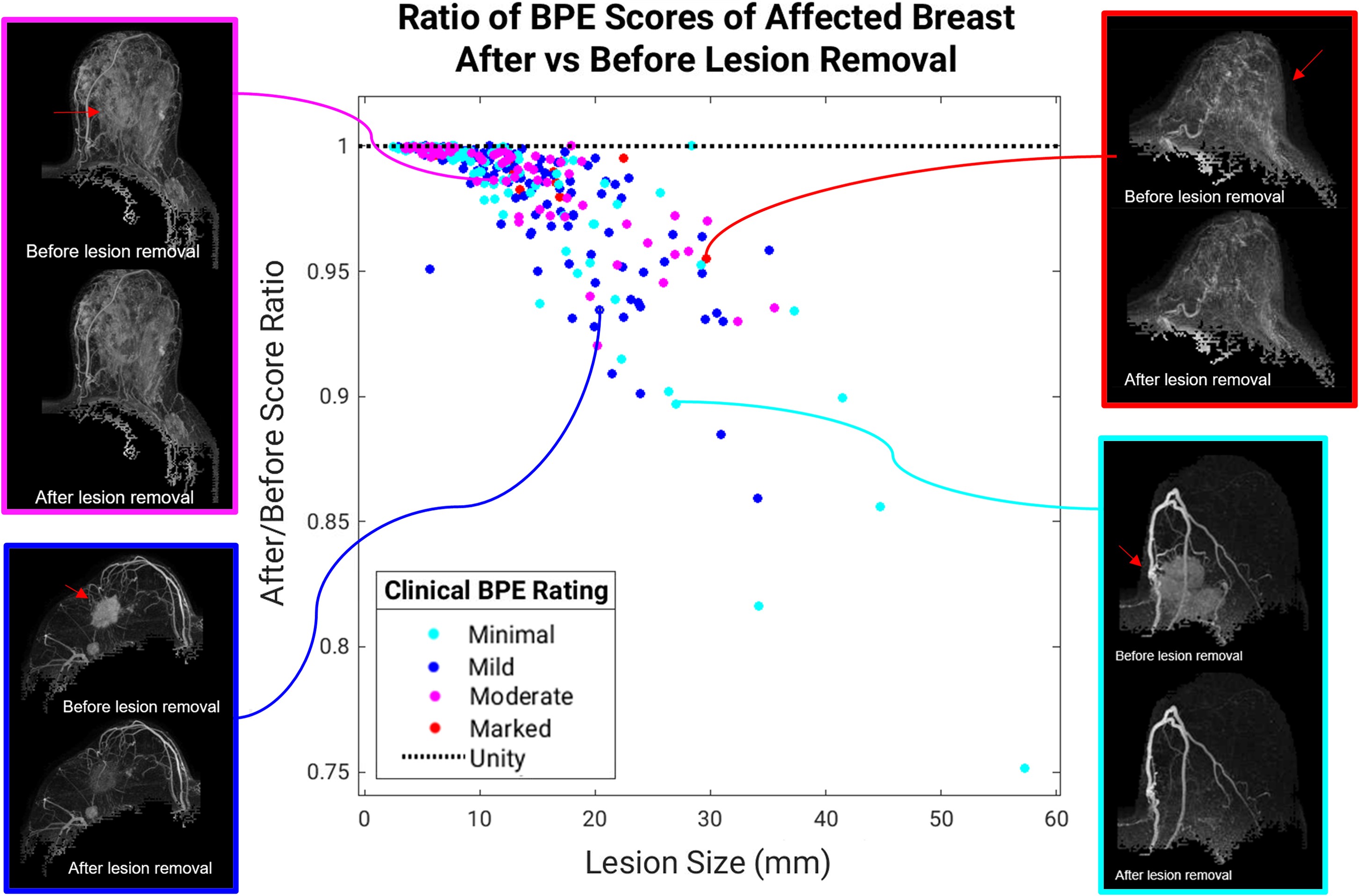
Ratio of the computer BPE score (second postcontrast subtraction MIP) calculated after lesion removal to the score calculated before lesion removal for the affected breast is shown versus the lesion size (n=350). Results demonstrate the importance of lesion removal to avoid inflation of computer BPE estimations, especially in cases containing large lesions and low BPE levels.
ADVANTAGES
ADVANTAGES
-
Enhances BPE quantification accuracy by digitally removing lesion-induced high-intensity signals
-
Reduces subjectivity and variability through automated, objective analysis
-
Improves diagnostic performance by more reliably differentiating between affected and unaffected breast tissue
-
Streamlines clinical workflows and integrates easily with existing imaging systems
APPLICATIONS
-
Breast MRI diagnostic automation
-
Cancer risk assessment software
-
AI imaging analysis tool
-
Clinical decision support system