Deep Learning-Based Diagnostic System For Differentiating Thyroid Tumor Genetic And Histologic Profiles
SUMMARY
A deep learning system analyzes high-resolution pathology images to predict thyroid tumor subtypes and gene expression patterns, helping distinguish less aggressive cancers from those requiring more intensive treatment
- The field of thyroid neoplasm diagnostics has long grappled with the inherent challenges of distinguishing between similar tumor subtypes using traditional histopathologic methods. The need for improved accuracy is underscored by the critical clinical decisions involved in differentiating indolent from aggressive thyroid forms, given that subtle morphological differences can lead to significantly different treatment strategies. This situation is compounded by evolving incidence patterns and the demand for more objective assessments that can keep pace with advances in medical science.
-
Current diagnostic approaches, however, are fraught with limitations. Conventional methods rely heavily on subjective interpretation and manual analysis, which often results in high interobserver variability and inconsistent results. Moreover, tissue sampling and imaging techniques sometimes suffer from issues like low resolution and background interference, which hinder the clear identification of nuanced features. The reliance on isolated histological or genomic markers without an integrated framework further compromises sensitivity and specificity, thereby limiting the reliability of existing diagnostic protocols in clinical practice.
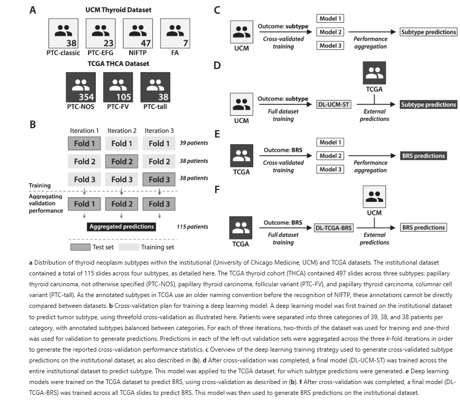
- The faculty inventor developed a deep learning system leverages an Xception-based architecture to analyze high-resolution pathology slides, converting them into standardized 299×299 pixel tiles. It processes images by removing background and incorporates pathologist-annotated regions to accurately capture diagnostic features. Two tailored deep learning models were developed: one predicts tumor subtype from an institutional dataset of thyroid neoplasms, while the other quantifies a continuous BRAF-RAS score from a 497-case The Cancer Genome Atlas cohort. This score reflects tumor gene expression similarity to either BRAFV600E or RAS-mutant patterns, aiding in the assessment of tumor behavior.
-
The technology is differentiated by its integration of genomic profiling with sophisticated image analysis, demonstrated by high sensitivities and specificities. The method’s ability to accurately distinguish between indolent and aggressive thyroid neoplasm subtypes—and map corresponding morphological variations—marks a significant advancement. Its quantitative BRAF-RAS score and mosaic mapping of tissue features provide unparalleled objective insight, potentially leading to improved diagnostic precision and optimized patient management.
FIGURE
ADVANTAGES
ADVANTAGES
-
Enhanced diagnostic accuracy with high sensitivity and specificity for distinguishing thyroid neoplasm subtypes
-
Objective integration of histologic features and genomic profiles via the BRAF-RAS score for precise tumor characterization
-
Facilitation of tailored treatment decisions by reliably identifying indolent NIFTPs versus more aggressive papillary thyroid carcinomas
-
Robust deep learning performance evidenced by strong statistical metrics (e.g., high AUC and R² values) that support clinical reliability
APPLICATIONS
- AI-enabled thyroid cancer diagnostics
- Pathology image analysis platform
- Genomic profiling integration tool
- Digital pathology workflow automation