AI-Based Ultrasound Analysis System For Automated Segmentation And Classification Of Adnexal Lesions
SUMMARY
AI-driven system automatically segments adnexal masses in ultrasound images, distinguishes different tissue components, and extracts key features to accurately classify lesions as benign or malignant, improving clinical diagnosis and workflow efficiency
- The field of medical imaging, particularly ultrasound-based assessment of adnexal masses, has long been recognized as critical for early detection and effective treatment planning in gynecologic oncology. Clinicians rely on ultrasound to noninvasively evaluate ovarian, fallopian tube, and surrounding tissue abnormalities. However, the inherent variability in imaging quality and the subtle presentation of tissue characteristics necessitate advanced solutions to enhance diagnostic accuracy while reducing operator dependency. This need stems from the increasing demand for precise, consistent assessments of adnexal lesions to better differentiate between benign and malignant conditions.
-
Current approaches to interpreting ultrasound images are fraught with challenges that impede accurate diagnosis. Manual segmentation and subjective interpretation introduce significant inter-observer variability, often leading to inconsistent lesion delineation and misclassification. Furthermore, conventional imaging techniques struggle to capture the nuanced differences in tissue composition, such as distinguishing between cystic and solid regions or detecting subtle pathological changes. These limitations result in diagnostic uncertainty and can delay the initiation of essential treatment, underscoring the urgency for improved methodologies in the evaluation of adnexal masses.
- The faculty inventor developed an approach employing a two-stage segmentation process where a deep learning U-net model initially isolates adnexal masses within ultrasound images, followed by an unsupervised fuzzy c-means clustering algorithm that divides the mass into distinct internal compartments. The system leverages expert-defined ground truths, data augmentation, and robust post-processing methods to ensure precise boundary delineation. It also extracts nine radiomic features related to morphology, geometry, and texture to support a linear discriminant analysis classifier, facilitating the differentiation between benign and malignant lesions with high predictive accuracy and minimal impact on clinical workflow.
-
What sets this method apart is its sophisticated compartmental analysis that discerns subtle internal variations such as cystic versus solid components, septations, and regions of necrosis or hemorrhage. By integrating both supervised and unsupervised techniques, it reduces human variability and enhances diagnostic confidence. Detailed performance metrics, including high Dice similarity coefficients and favorable Hausdorff distance ratios, underscore its reliability, offering a nuanced, highly efficient tool for more accurate clinical decision making.
FIGURE
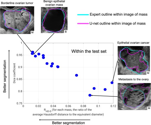
U-net segmentation performance in the test set in the task of segmenting the entire adnexal mass from the surrounding tissue, compared to expert outlines. (RHD-D: ratio of the average Hausdorff distance to the effective diameter of the mass.) Images of the four masses with the best performance (highest Dice coefficient and lowest RHD-D) and lowest performance (lowest Dice coefficient and highest RHD-D) are shown. Clockwise from top left with pathology (with patient diagnosis) details: borderline ovarian mass (borderline serous tumor), epithelial ovarian mass (benign serous cystadenoma), epithelial ovarian cancer (high-grade serous ovarian cancer), and metastasis to the ovaries (cancer of gastro-intestinal primary origin). B, benign and M, malignant.
ADVANTAGES
ADVANTAGES
-
High diagnostic accuracy with robust segmentation results and classification performance (e.g., high Dice coefficients and AUC scores)
-
Reduced human variability by automating lesion segmentation and feature extraction, ensuring consistency
-
Detailed analysis of tumor characteristics by compartmentalizing lesions into cystic and solid regions
-
Reliable malignancy screening with a high negative predictive value to confidently rule out cancer
-
Minimal clinical workflow disruption with a simple bounding-box input and automated processing integration
APPLICATIONS
- AI ultrasound diagnosis
- Ovarian tumor detection
- Automated lesion segmentation
- Radiomic risk stratification
- Clinical workflow automation
- Women's health